
Nemanja Rajković,1, Bojana Krstonošić,2, Nebojša Milošević,1
1 Department of Biophysics, School of Medicine, University of Belgrade, Višegradska 26/2, 11000 Belgrade, Serbia; 2 Department of Anatomy, School of Medicine, University of Novi Sad, Hajduk Veljkova 21, 21000 Novi Sad, Serbia.
DOI: 10.1155/2017/8967902
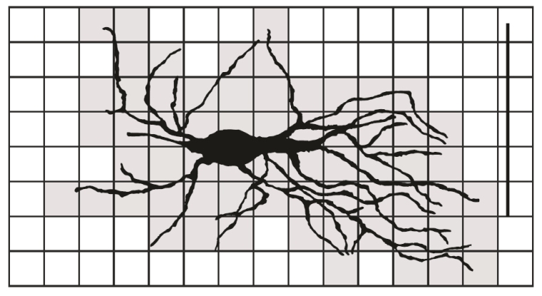
This study calls attention to the difference between traditional box-counting method and its modification. The appropriate scaling factor, influence on image size and resolution, and image rotation, as well as different image presentation, are showed on the sample of asymmetrical neurons from the monkey dentate nucleus. The standard BC method and its modification were evaluated on the sample of 2D neuronal images from the human neostriatum. In addition, three box dimensions (which estimate the space-filling property, the shape, complexity, and the irregularity of dendritic tree) were used to evaluate differences in the morphology of type III aspiny neurons between two parts of the neostriatum.
